With the scientific analysis of works of art becoming more and more sought after, the author, after briefly referring to various specialized laboratories, explains how to find the most competent laboratory best able to analyse a work of art and raises the issue of the reliability of the analyses produced. He then sets out a brief history of scientific examinations and studies the various techniques used, which he summarizes in table form: examinations and analyses of surfaces, examinations and analyses of internal structures, dating methods. He ends his liberally illustrated article with the issue of the probative value of analyses carried out by scientific police laboratories.
La revue Experts n° 19 – 06/1993 © Revue Experts
After a vertiginous ascension of the art market and its abrupt collapse, investors query the value of their investment.
Too many artist attributions have changed and a certificate from one or two accredited experts for a painting that exceeds one million francs is no longer sufficient. The banks, which have not been spared this crisis, demand an increasing number of expert reports and scientific guarantees to cover their involvement. The same applies to insurance companies, which, inundated with disputes, revise their contracts and, more and more frequently, turn to scientific examinations and marking. Auctioneers suffer losses, antique dealers attempt to sell their stock and reduce costs awaiting a hypothetical market recovery. In this generalized recession, certain attempt to distinguish themselves by offering their clients better quality goods, accompanied by full historical and scientific information.
Science, therefore, has an increasingly role in the analyzing of works of art.
The various laboratories in France
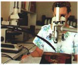
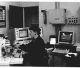
Measuring annual growth rings on an oak panel with the aid of a binocular loupe and an automatic placement table linked to a computer.
Photo G. P. Lab.
A number of large State laboratories exist in France, such as the Laboratoire de Recherche des Musées de France (L.R.M.F.) [French Museums Research Laboratory], the Laboratoire de Recherche des Monuments Historiques (L.R.M.H.) [Historical Monuments Research Laboratory] or the Laboratoire de la Police Scientifique in Paris [Paris Scientific Police Laboratory]. Unfortunately, because their purpose is to examine public collections or classified or controlled works, it is difficult for individuals to have access to their services.
Private laboratories that specialize in the study of works of art that themselves carry out such examinations using the appropriate equipment and personnel can present two advantages: rapidity and independence. Rapidity because most analyses are carried out in less than one month, encouraging civil servants to use their services. Independence because they are exempt from demanding administrative procedures and can better adapt to changes in technology, except in relation to extremely expensive equipment.
Nowadays, numerous universities or European organization restorers such as IRPA, IFROA, ICCROM or Restauro are becoming art consultants or even ‘analysis laboratories’. These are small organizations that are better equipped to carry out x-rays, microscopic, ultraviolet and infrared analyses. If they themselves are not able to carry out certain delicate examinations, they subcontract out or orient their clients to laboratories with more sophisticated equipment.
How can you determine which is the most competent and best able to analyse a work of art?
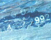
It is primarily used on works of art where a person wants to engrave an unalterable text, visible only by microscope, to the work. Reading the marking is instantaneous and does not involve technical costs.
The laboratory can quote for marking the pictorial layer of paintings and bronzes.
A competent laboratory will carry out most of the examinations itself. It will require that the work of art be left in its possession, without any specific time limit (between one week and one month) in order to check the results, if necessary, and determine, providing full guarantees, which areas to analyse.
It must comprise a team of scientists assisted by restorers and curators, the former operating sophisticated devices with ease and rigour, the latter guiding the research, which depends on the history of the work in question and the materials and techniques used to create it.
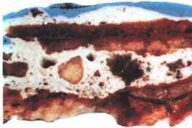
For example, if a scientist uses a microscope to analyse the cross-section of a multi-coloured sculpture that has, since the Middle Ages, been repainted several times, he risks confusing the preparatory layers, the paint and any special effects such as glazing or applied brocade*. If he does not have any experience in ancient techniques or if he is not assisted by a restorer or curator, he might interpret each layer as a visible layer. In the past, certain studies have claimed that gold-plated virgins were yellow and red ochre, whereas, in fact these were intermediary layers between the preparation and the gold leaf.
In another example, an analysis laboratory made the news in the 1980s when analyzing pigments in a restored part of a 17th-century painting. The conclusion created a scandal and a counter-expert report proved the authenticity of the painting and, as a result, the incompetence of the scientists who had been brought in without specialized technical assistance. A laboratory that analyses works of art must combine scientists, historians and restorers. Without such close collaboration between the three parties, significant errors can occur.
* Stamps in wax, gilded, glued to clothes, on statues and paintings in the 15th and the beginning of the 16th centuries to represent fabrics woven with gold thread.
The reliability of analyses, science and conscience
Scientific results are, a priori, reliable and irrevocable. But experience shows us that there always remains a risk, even minimal, of a poor interpretation through lack of experience, concentration, time or the desire to rapidly finish the analysis…
The conscience (ethics) requires each analysis to be checked and all results returned to the interested parties. The results must be clear and illustrated with photographs taken during the various examinations. They must also contain all results from advanced tests with localization of the examinations, the elements analysed and the corresponding spectrums. So that any expert or judge, assisted by a scientist, can assess the veracity of the examinations.
In certain circumstances, the scientist should not fear expressing doubts where an uncertainty cannot be removed.
A good scientist is not the person who knows everything. Admitting to a client or a judge that it is too risky to come to a conclusion is not always easy because indecision slows the whole process down. Those who know everything are reassuring and can speed things up. Neither should the scientist fall into the reverse situation where he prefers declaring an atypical painting to be a fake rather than take risks.
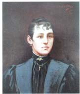
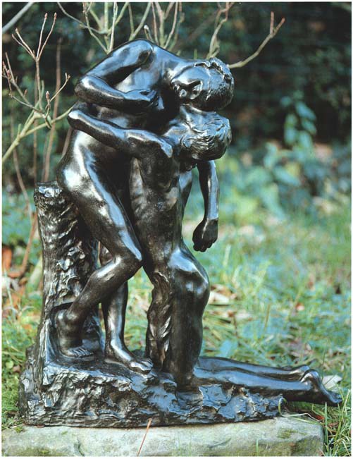
The work of Camille Claudel is very limited. To protect herself against fraud, Mrs Reine Marie Paris, great-niece of the artist, rights holder and expert in her work, frequently consults the Versailles Analysis Laboratory to create a database on the technique and composition of sculptures in bronze and paintings.
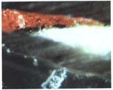
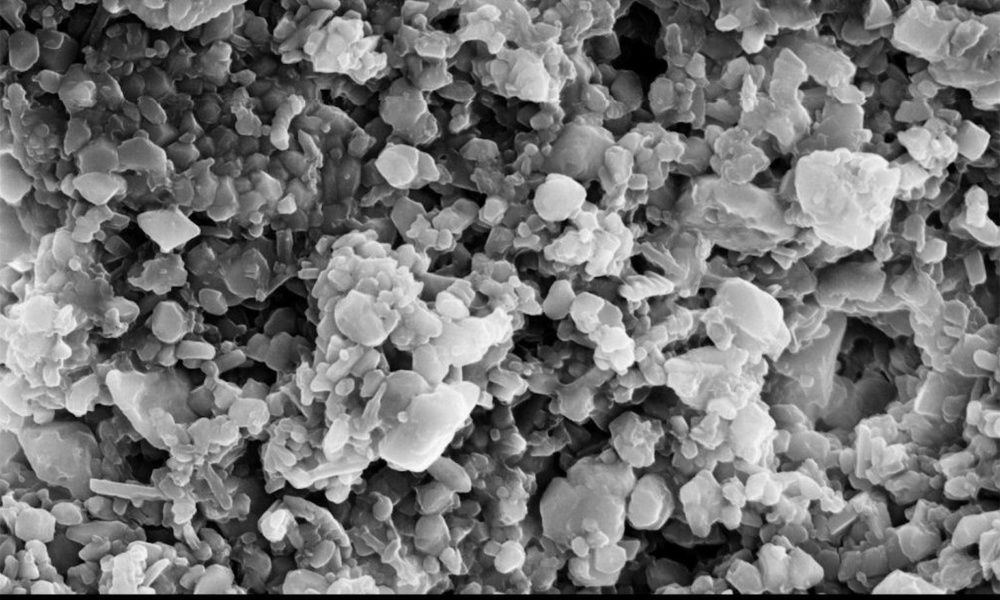
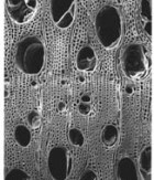
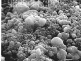
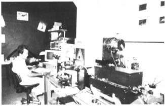
The operator localizes the coloured grain that he will analyze using a scanning electron microscope. The white layer corresponds to the preparation of the canvas.
The view is monochrome because the secondary electrons forming the image do not transmit the colours.
A brief history of scientific examinations
The palette of scientific examinations has increased over the years. Radiographies and observations under microscopes were initially used in the 1950s and the following decade saw the growth in the use of ultraviolet and infrared light. It was particularly in the 1970s, due to computers, that new techniques developed for dating, carbon-14 and dendrochronology; for the study of pigments, the scanning electron microscopy, X-ray micro-fluorescence X, spectrometry, etc.
Lastly, new technologies were developed in the 1980s such as tomography (scanner), nuclear magnetic resonance, the Louvre (AGLAE) and the French Museums Research Laboratory (L.R.M.F.) particle accelerators and the Raman Fluorescence Absorption Reflection (RAFLAR) at the Scientific Police Laboratory.
All of these examinations and analyses are for the purpose of studying the materials and techniques used in producing works of art.
Such examinations provide information on the history of the object, the manner and era in which it was produced. Its current condition can be determined as well as the methods that will be best adapted to restore it if it has suffered significant damage.
The scientific approach is, in fact, quite simple. It uses non-destructive methods or takes microscopic samples, examining both the surfaces and what is invisible.
What is the difference between an examination and an analysis?
L’examen est le résultat d’une observation d’un objet, tandis que l’analyse est l’identification des composants. Nous présentons ci-après un classement sommaire des examens et des analyses dont la définition est donnée précisément dans le tableau inspiré des documents du Laboratoire de Recherche des Musées de France.
The observation techniques used and their applications

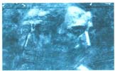
The first examination carried out is, obviously, a visual examination of the work using direct or angled lighting that identifies, for example, the level of flaking in the pictorial layer of a painting. True professionals commonly carry out analyses using strong magnifying glasses or optical microscopes under either natural, filtered or angled lighting or the naked eye using ultraviolet or infrared lighting.
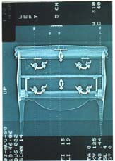
Radiography, scans, tomographic cross-sections and nuclear magnetic resonance enable an observation of the interior of objects and certain conclusions to be drawn. From the radiography of a painting, it is easy to discern whether it has been painted over, whether there is a hidden signature or an expertly hidden restoration. A scanner may determine the authenticity of all of the aspects of a sculpture of the Virgin Mary or an item of furniture. Nuclear magnetic resonance perfectly localizes xylophage insect larvae or any variations in the humidity of wood.
The techniques used to analyse components
There are still numerous experts who refuse to analyse the components of a painting. Sooner or later, however, they will discover that such analyses, which relate to the binding agents and pigments, provide precious information.
They can filter out most counterfeits using the ‘on the contrary’ principle of proof. They cannot, however, by themselves but with rare exceptions, determine if the painting is by a master painter, his workshop assistants or a counterfeiter who was a contemporary of the presumed artist.
The most commonly used techniques are the pigment analyses using a scanning electron microscope (SEM) or x-ray fluorescence (a more rapid but less precise technique), by microchemistry using reacting agents on micro-samples or spectrometry.
Other methods are still the subject of research, in particular the measuring of the natural radioactivity of lead that enables lead white to be dated ± 20 years.
Binding agent analysis has developed over the past five years, giving rise to the possibility of defining relatively short, precise, periods for 20th-century paintings.
What, for example, is the conclusion to be drawn on a painting that had a binding agent, acrylic, that was used only after the 1960s and the artist, who died ten years previously, had no idea of the existence of acrylics! While science cannot be formal in attributing origin, it can, on the other hand, be formal when disavowing paternity.
Dating techniques
Thermoluminescence, carbon-14 recording and dendrochronology are dating techniques that are appropriate for certain materials.
Thermoluminescence only works on earthenware. When heated to more than 700 °C, the sample will emit rays that will be proportionate to the number of years that have passed since it was produced. Its response curve has a margin of error of a hundred or so years (precision coefficient of between 5 and 15%).
Despite such uncertainty, antique earthenware is no longer sold in major foreign sales without its thermoluminescence dating certificate.
Carbon-14 dating enables the age of organic objects that are between 2,000 and 30,000 years old to be defined to within a precision of ± 200 years. This method is, therefore, used only for antique and prehistoric
objects. It consists of measuring the natural radioactivity of a material. It has been found that carbon 14, diluted by the abundance of carbon 12 produced by factories and coal-fired stoves, was reduced by one-half at the end of the 19th century. Then its presence abruptly increased, doubling since 1900. After the Second World War, it became radioactive. For the past 10 years, researchers have been attempting to detect such phases in linseed oil used in paintings in order to facilitate their dating by placing their age in three main periods – before 1900, from 1900 to 1945 and from 1945 to today. It is likely that such work will be successful within the next five years and will substantially affect attributions of origin.
Dendrochronology is, on the other hand, very precise as it dates fragments of wood to a specific year by measuring the extent of the spring and summer tree rings, following by comparing them to a scale of reference. However, this science can be applied only to certain woods such as oak and some softwoods. It is, nonetheless, very precious when carrying out expert reports on Dutch and German paintings that, until the 17th century, were nearly all painted on oak.
All of these forms of analysis, which are not exhaustive, provide precious information on the art object. Applied and interpreted by competent people, they help prevent counterfeiting and improve the knowledge of art historians, sometimes even enabling the discovery of paintings hidden in attics.
Such techniques should be grouped according to the results sought: surface examinations, internal structure examinations, surface analyses, internal structure analyses, dating of materials.
Do the analyses carried out by the Police Laboratory constitute an irrefutable expert certificate?
The idea of creating a specialized identification unit dates back to 1883 when, for the first time, the police used the Bertillon anthropometric method to identify a recidivist.
Since then, the service has grown considerably and, in 1985, became the Paris Scientific Police Laboratory (L.P.S.). Established in the heart of the courthouse in the Ile de la Cité, it has four branch offices in Lille, Lyon, Marseille and Toulouse and employs 19 scientists, nine administration assistants and 23 police officers.
Used both nationally and internationally, it operates on request by judges in either criminal or civil proceedings (principally where the two parties, ie, the plaintiff and the defendant, have opposing views) or when requested by an individual.
An individual who wishes to have his object analysed by the Scientific Police Laboratory must commence ex parte proceedings (where there is no dispute, as opposed to inter partes) in the Tribunal de Grande Instance (Regional Court) where he lives. With the judicial system being saturated, only applications relating to exceptional objects have any chance of success.
The results of the analyses are not an ‘expert certificate’ but help the judicial system produce a solid body of evidence that orients the conclusions drawn by experts.
In this search for evidence to enable a decision one way or the other, the laboratory will give priority to non-destructive methods and samples will be taken only as a last resort. It should be remembered that the work being analysed is, after all, an item of evidence!
First, the standard methods of observation will be used, such as radiography, infrared photography, ultraviolet waves … and the RAFLAR. This device enables microanalysis without damaging the work and the identification of all of the pigments used. It is, therefore, easy compare the results obtained with a table tracing the development of pigments and techniques. Anachronisms in the production of the work can, therefore, be identified.
This method is very interesting for synthetic pigments, the dates of fabrication of which can be precisely defined as being between the middle of the 19th century and the 1950s. The white pigment titanium dioxide, for example, exists in two forms, Anatase and Rutile. The first was commercialized in France in 1925 and the second appeared in the American market in 1941 and in Europe after the Second World War.
If the non-destructive methods do not produce any results, the analysis of pigments will be carried out by taking a sample, which will be examined using a scanning electron microscope, x-ray fluorescence or diffraction.
It should be remembered, however, that, in certain cases, the scientific examination remains insufficient in determining whether the object is a counterfeit or not. This is the reason why art experts and, more particular, experts in paintings, include not only the scientific analysis but also apply an artistic approach to the painting.
Examination and analysis techniques
Surface examinations
Technique |
Process |
Applications |
Binocular magnifier |
Stereoscopic view showing relief with enlargements from 1 to 100. Lighting is often external and directional using optical fibres. | Condition: alteration (corrosion, restoration, cracks). Fabrication techniques: assembly, finalizing, tool cuts. Surface condition: blisters, inclusions, colouring agents, homogeneity or heterogeneity. |
Optical microscope |
Enlarged view of the surface or internally using a microscopic sample, enlargement from 100 to 1,000. | Observations of the surface of paintings, wood, metals, stone in order to assess its porosity, damage. |
Oblique or low light |
A directional luminous source oriented according to the surface plane improves visibility of the relief by highlighting shadows. | Study of the traces left by fabrication tools. Discovering artisanal restorations, fakes… Stone, granulometry. Iron, hammering. Inscriptions in hollows. Cracks, surface deformations. |
Fluorescence under UV lighting |
A light source using mercury gas that emits UV rays, provoking fluorescence. | The UV rays localize the heterogeneity of the lighted surface and show restorations (commonly used to analyse paintings but also cabinetmaker varnishes). They can differentiate certain materials such as: lacquer, antique crimson, ruby and garnet. |
Photographs and infrared reflectography |
Certain substances are more visible under close IR rays than by eye. Such rays enable the layers underneath the surface skin of the material to be seen on photo film or a screen monitor. | Reading effaced marks and inscriptions on parchment, ceramics, wood, stone, paint and canvas. |
Examinations of internal structures
Technique |
Process |
Applications |
Macro and micro-endoscopy |
Frontal, lateral and oblique observation through the introduction or flexible or rigid optical fibres inside an object. | Study of the techniques used to fabricate hollow objects. Bronzes: presence of core, localisation of struts, inserts… Wood: assembly method Ceramics: splits, streaks. Iron: oxidation state Stone: Condition of cracks, joints… |
X-rays |
The length of the x-ray wave conditions its penetration. The absorption depends on the nature of the materials, their volume, density and atomic number. X-rays that have traversed the object are recorded in white on a film. | Painting: Pictorial technique (initial sketch, repainting, change of composition, overlayering, overpainting). Condition of the base media and the pictorial layer. Identification of various parts of an object Fabrication techniques General condition |
Tomography (scanner) |
– Non-metallic objects – The object is placed on a mobile table. An x-ray tube pivots around it and produces close (overlapping) or distant cross-sections. The results are recorded in the computer, which then rebuilds, from the digital information, an image in 2 or 3 dimensions. |
Very localized internal observations. Reconstruction of absent parts or those to be removed. Non-destructive internal view that enables, among other things, the discovering of added parts not visible from the exterior. |
Analyses of surfaces
Technique |
Process |
Applications |
Micro-fluorescence X |
Superficial charging of the air by an x-ray, the material emits a secondary x-ray that contains the characteristics of its elementary composition. The analyses are one-off, non-destructive, immediate and can be read on a spectrum produced by the computer. |
Analyses of the surface of all materials, organic materials, glass, pigments, etc. |
(1) Visible and (2) UV (RAFLAR) spectrometry |
Charging of the air by an electrical discharge (1) or by a laser ray (2), the surface layer of a metal emits a spectrum that contains the characteristics of its elementary composition. The analyses are one-off, panoramic, immediate. | Analyses of any form or dimension of metal to millimetric levels. Reveals the composition of alloys, soldered joints, etc. |
Analyses of internal structures
Technique |
Process |
Applications |
Infrared absorption spectrometry |
Charged by a continuous spectrum, the inter-atomic bonds of a molecule selectively absorb energy photons equivalent to the energy transitions of vibrations. | Identification of organic bonding agents (waxes, resins, polymers), inorganic components, carbonates, sulfates, the results of corrosion. |
Scanning electron microscopy |
Charged in a vacuum by electrons, the material emits an x-ray that enables micrometric identification of the elementary composition. | Identification of all elements, even in the form of traces. Study of sources, conservation, authentication. |
Dating methods
Technique |
Process |
Applications |
Dendrochronology |
Wood dating through the observation of annual tree rings. Trees that are subject to climatic conditions in a particular region produce an identical result in the width of the tree rings. Recorded, therefore, are ring sequences that enable dating by way of comparison with reference materials. |
Dating of certain woods. |
Carbon 14 |
When an organism dies, the carbon 14 contained in it starts to decay, while its carbon 12 remains stable. One-half of carbon 14 disappears every 5,568 years. |
Dating of organic items, antique woods, charcoals, tissues, etc.. |
Thermoluminescence |
The natural radioactive elements (uranium, thorium and potassium) present in rocks modify the energy levels of electrons. By heating them to 500 °C, the electrons release energy in the form of light, which is then measured. Following firing, a ceramic accumulates a dose through natural irradiation. Refiring a powdered sample in a laboratory enables the duration of the irradiation to be measured from the quantity of light emitted. |
Dating of earthenware, tiles, ceramics, clay moulds for hollow bronzes, subject to certain reservations. |
Published with kind permission from the L’Estampille/L’Objet d’Art review. Extract from no. 268.