The author reveals the first results that he has obtained using a scanner to detect fake objects in the art world. Following an examination of painted wooden statues and tree rings (to determine growth), the author discusses new investigative methods: the identification of wood, reconstitutions and silver plating, the direction of metal inserts, observing the progression of xylophage insects and treatments, the study of how furniture is assembled, musical instruments, observing paintings and detecting restorations to vitreous glass.
Revue Experts n° 11 – 06/1991 © Revue Experts
Often skilfully executed and difficult to detect in a preliminary examination, fakes are a challenge for experts. Scientific methods of analysis are an indispensable aid. The use of scanners, until now used only for medical patients, will undoubtedly, in light of the first examples presented in this article, be a precious tool in distinguishing fakes from real works of art.
The soaring prices of works of art, due mainly to demand being greater than supply and with interest remaining substantial, more than ever attract counterfeiters and fakes are multiplying in the market.
What remedies do experts have when faced with this chronic virus? First, substantial experience, a keen sense of observation of the smallest details and a perfect knowledge of the techniques used to produce the object the subject of an expert examination. This is not, however, sufficient in today’s world. Well-known counterfeiters hide others who have resisted the temptation to show themselves. How many experts are there who rely only on their flair but are clearly mistaken? The lesson can be harsh for many, who have been forced to admit their limits.
The incessant and increasing afflux of new works, with keen interest in Art Deco, art from the 1950s and modern art, does not facilitate the work of expert firms, which have, as a result, increased in number. The ensuing competition, with ridiculously low rates being applied – from 1% of the value of the object being examined (from FF 0 to 50,000) – does not permit a serious examination of the work. Such basic expert valuations are, in fact, often merely brief, intuitive, assessments by the expert who visually compares a signature, a pleat or the curve of the foot of a chest of drawers when giving his advice.
Grouped expert assessments for an inventory or before an auction produce variable results and can contain surprises as it is difficult to assess 200 objects in two or three hours without making mistakes or being fooled by some.
An intuitive assessment provides initial guidance but can no longer be used for a sale of a work of art that is worth thousands if not millions of francs, the value of which attracts fakers.
Serious professional no longer offer an important object for sale without a full accompanying file attesting to its origins and authenticity. Numerous disputes between experts in well-known proceedings have also convinced the courts that assessments by experts should no longer be based solely on the inner conviction of the professional and that proof in the form of a photo or an analysis report carries more weight than reputation.
Fakes – a chronic virus that needs to be eradicated
Modern analysis techniques, the scope of application of which does not cease to expand, have also become more refined and enable the expert to base his conclusions on irrefutable proof. That said, however, as in medicine, the examinations or x-rays only produce what the operator seeks. Aspects like the reliability of analyses, the judicious choice of samples and the quality of the photographs are of primordial importance.
Science can turn a lot of preconceived ideas on their heads. Works of art seen to be authentic over several generations can suddenly fall from their pedestal following a scientific examination.
In the current transitional period, certain professionals attempt to ignore such recent advances, claiming they are not necessary. But what should we do with all those objects that have been sold several times at auction, with certificates, the authenticity of which is challenged by a museum during purchase negotiations? Current events provide us with a reminder, for example the case of a presumed Rubens’ Study of the Head of Saint Jerome. This painting was auctioned on 20 March 1956 by the auctioneer Maurice Rheims, assisted by his expert, guaranteeing its authenticity, was subsequently auctioned several times but was declared to be a fake by Mr Foucart, the Head Curator of Paintings at the Louvre Museum. Mr Foucart included in his report, among other things, a micro x-ray fluorescence scan carried out by the Research Laboratory of the Museums of France, which highlighted a significant proportion of zinc in the yellow tints. This discovery constituted irrefutable proof of a dating error, because yellow with zinc only appeared at the beginning of the 19th century.
Curators, who take every precaution, always produce a full analysis before deciding on any acquisition.
There are many private individuals who have suffered the stinging affront of a refusal when proposing a presumably important work as a gift or donation.
Science serving expert reports: an impressive arsenal
After radiography, used from the 1970s by the Laboratory of the Museums of France, other cutting-edge techniques used, in particular, in medicine, have been applied in examining works of art.
Observations under microscope, with photographic shots and angled lighting and using fibre optics have become generalized. Analyses of the elements constituting a painting, through the application of reactive agents on micro samples or by applying various ionic rays (the A.G.L.A.E. particle accelerator at the Research Laboratory of the Museums of France), microprobes or scanning electron microscopes have become commonplace, as have the sciences determining the age of materials such as the carbon-14 dating of archaeological objects, thermoluminescence of antique earthenware or dendrochronology (ring counting) for certain woods.
All of these examples, used only in the last 20 years, are easily accessible to private individuals. Only access to the A.G.L.A.E. machine requires long negotiations with the State. In the past two years, our laboratory offers its services to national museums, classified and supervised museums and private individuals and our success has led us to branch out into such diverse areas as the identification of gilding on metals, pigments, the analysis of objects using x-rays. The first results are so spectacular that it would seem appropriate that we disclose them to the wider public.
A new investigation technique: x-ray scanning
A CT or ‘computed tomography’ scan of wood, tissue, limestone, etc, provides additional information to that obtained from radiography. The observations obtained by the computer are selected on a screen and sent by the operator to a film negative. The very precise images constitute irrefutable proof that leave no room for controversy. The image produced by the computer is so close to reality that, compared to radiographies, a neophyte can assess its full impact.
In brief, the scanner comprises five separate elements: a mobile tube that emits x-rays, a battery of detectors opposite it, a computer that carries out the calculations, a command console with visualisation screens and a device to produce and print photographs.
The object being observed is placed in the centre of the tube on a moving table that is controlled to the nearest millimetre by the operator. This precision offers the possibility of measuring, among other things, the internal dimensions as seen on the scanned image and the distance that separates each cross-section.
The first process carried out is an overall scan or ‘scout view’, ie, a digital image of the overall object either from the front or the side. This image is similar to that of an x-ray but is more precise and enables the operator to find areas of interest and those that contain undesirable objects. Cross-section lines are then defined and programmed. The computer then receives all of the cross-section scans, which have approximately 2,000 shades of grey, and transfers the variations in density in the object being examined onto film.
 |
|
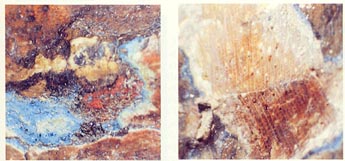
| An initial scan shows the volumes and metal inserts. Markings for the cross-sections – the dotted lines in the photo – are then positioned. | The lower portion of the scan reveals that the sculpture is fixed to the base by four very ancient nails that are rusty, broken and invisible from the surface. |
The first applications in the examination of works of art: painted wooden statues
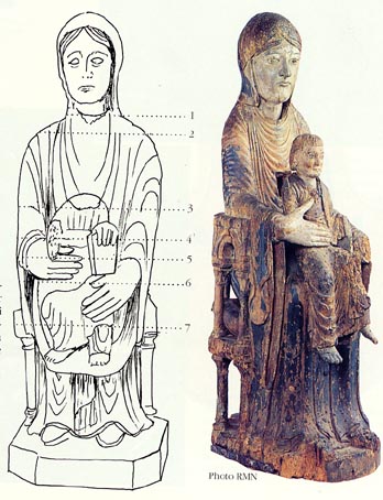
Where a wooden sculpture is entirely covered by gilding, plaster or paint, the question that is often posed is whether the statue is homogenous or not, ie, does it consist of a single piece and only one type of wood? The issue arose for us in relation to a wooden Virgin and Child from the 13th century that appeared to be entirely from that era.
An initial examination under a microscope showed us the presence of joints under the paint, confirmed by an abrupt change in the orientation of the wood grain. These observations were able to be made, however, only in areas that lacked paint, which prevented us from following the glued joints and exhaustively locating all of the external pieces.
 |
| Scanned cross-section n°4 localises the area attacked by xylophage insects and show that a substantial part of the sculpture under the paint remains unaffected |
It should be noted that a vertical x-ray would have provided some interesting information on, in particular, the addition of the head of the Virgin, the existence of two dowels in the joint and the absence of any hidden cavity that might hide religious relics or other precious objects. It would not have been possible to obtain any details on the pieces of wood that had been added.
CT scan cross-sections were therefore taken in various places determined by the initial scan in order to refine the microscope analysis.
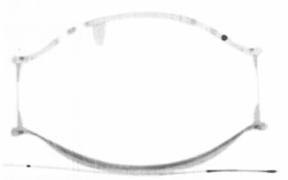
An image was taken of the neck exactly at the site of the two dowels to determine the nature and condition of each. The scan, which reconstitutes an image that is perpendicular to the grain of the wood, enables the annual rings in the homogenous wood to be very clearly distinguished and facilitates the identification of the wood used.
One of the dowels is, like the statue itself, made of walnut wood and is clearly in poor condition. The second, on the other hand, has the ring structure of a resinous wood and has not been attacked by xylophage insects, which suggests that it is more recent than the first and that it is, therefore, a subsequent consolidation.
The second scan shows the penetration of the dowels but still does not show any presumed cavities.
Splits visible from the exterior suggest that the Child Jesus was separated from the Virgin. The scan of this area shows that, in fact, the woods used are in perfect continuity and, especially, are identically orientated. This cross-section clearly shows that the sculpture was carved from a single piece of wood. The poor condition of a peripheral ring, which is similar to sapwood, fully confirms this.
The final cross-section, near the seat of the chair, highlights a large number of small pieces of wood identifiable not only due to their joints but also the differences in the orientation of their rings.
All are found in the peripheral aspects of the sculpture and are in excellent condition, even though they were glued to areas in very poor condition.
These relatively recent restorations, which suffer from varying conditions of deterioration, do not all appear on the surface and only a CT scan can find them.
In these circumstances, an examination of the paint also enables a better assessment of the thickness of the plaster (or preparatory layer) that is heavy in limestone (calcium carbonate), which can be clearly seen in the image. The more recent additions in oil do not appear in the view chosen.
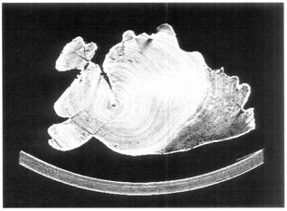
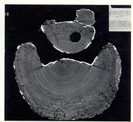
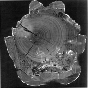
Observation of annual rings and ring counting
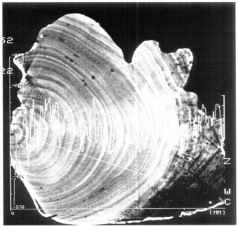 |
| Compared with usual ring-counting method, the scanner enables an internal and very precise localisation of the wood rings. This image shows the density the variations from the line of the right of the heart of the wood ring attacked by insects, showing the exact variations in the density of each annual ring and their width. The time spent in dating the wood will thus be considerably reduced. |
One of the interesting observations from CT scans, the orientation of wood rings, has been evoked on several occasions. A radiograph also provides an image of wood rings but on the condition of being exactly positioned on the traversal axis and must be used on a thickness of wood that is relatively small in order to clearly identify the ring deviations. For CT scans, the traversal reconstitution is, on the contrary, automatic and is localised very precisely in the object.
This technique is, therefore, of value for counting wood rings in objects that do not have a cross-section that is easily accessible or that is too large in size.
This is often the case with sculptures, where the base is hidden by a support or has larger dimensions than those that might be the subject of a possible scan through the object at, for example, waist level or the arms of the Child Jesus. Using a CT scan, a greater number of measurements can be taken, at the same time providing support for the dating. Since our work began, our research has included CT scans in order to provide greater detail on the ring counting. The computer offers the possibility of analysing the density of the annual rings and reproducing them either in a scanned image or in the form of a graph that is very similar to that used in ring counting.
Futures investigations
After only several months of research, new perspectives have arisen:
The identification of the wood used. A sample table produced using the computer processing of numerous samples will soon be completed for commonly used woods that will make it easier to compare woods that is not possible by the naked eye.
The reconstitution of a volume. Due to computers, the CT scan cross-sections taken side-by-side or overlapping offer the possibility of reconstructing an object in three dimensions and then extracting different types of plans from it. Simulations of volumes are also possible and even elements that no longer exist can be reconstituted or a copy made using data from a computer-directed pantograph.
These techniques are common in the United States for the creation of bone prostheses. In France, researchers use this process to reproduce natural-looking busts in expanded polystyrene or other synthetic resin of low density.
The detection of gold and silver plating. The detection of gold and silver plating hidden under successive layers of paint is a common problem.
By choosing a precise and appropriate window, the areas of gold or silver plating can be highlighted even though the gold leaves are less than 4 to 8 microns thick. While the metal can be easily localised, our techniques are not sufficient advanced to enable their nature to be precisely defined.
The detection of metal inserts. Due to the scanner, we can localise, to within half a millimetre, the position of screws, nails and any other hidden metal insert. Clearly, this level of precision facilitates the removal of such objects and their identification, eg, whether the nails are modern or ancient. Due to their substantial density compared to wood, metal inserts are artefacts that adversely affect the CT scan, the operator therefore being careful to programme the cross-sections to avoid them if he wants to obtain a clear image of the wood.
The detection of metal inserts is also required when we want to analyse objects using nuclear magnetic resonance.
Observing the progression of xylophage insects and treatments. Wood that is seen from the outside to be excessively worm-eaten may not necessarily be worm-eaten to a great depth. Xylophage insects often restrict their activity to a humid area such as the back of a statue that touches a wall or the underside of a wooden panel. The CT scan cross-sections can very precisely define the extent of their activity.
The most common damage to works of art in France are caused by lyctus and furniture beetles, followed by the capricorn beetle and termites, the galleries of which are easily identifiable using the scanner. CT scan cross-sections can, in most cases, tell us if the insects are still inside the object and, in some cases, determine if the larvae continue to progress.
These observations also enable treatment using a liquid such as ‘xylophene’ and determine its penetration and effectiveness. The same observations are possible after impregnation by consolidating resins such as Paraloid or epoxies.
Studying how furniture is assembled. The diameter of the tube is currently limited to 70 cm with a reduced ‘vision’ of 50 cm, which excludes an overall view by the scanner. Small items of furniture, however, such as decorated works by master craftsmen, dolls, which we have previously studied, chairs and certain average sized items such as chests of drawers can, for larger items, reveal interesting information from a tangential cross-section of part of the object.
Assemblies are examined without being taken apart to determine the techniques used at the time the object was built or if it has been restored or if it is in perfect condition. Hasty and irreversible restorative work is also localised to avoid risky quotes by restorers who will no longer be hindered by restorations packed with epoxy glues such as Araldite, which was very popular in the 1960s and 70s.
Studying musical instruments. The examination of musical instruments by CT scan enables not only the visualisation and reproduction of the sinuous curves of the instrument and the rapid identification of essential features but also the thickness of the materials used and the method by which it was assembled. Certain thicknesses that are unable to be measured due to the difficulty of access are taken in a few seconds, stored and reproduced! Experts will see that this is a precious tool in establishing their conclusions and supporting their statements.
Undoubtedly, there is an immense scope of applications in this area, which can be further developed to better understand the sonority of instruments and the secrets of the master craftsmen.
Examining paintings. While a scanned image offers a good overall ‘x-rayed’ vision of an oil on canvas, wood or other material and enables paint preparations or layers to be distinguished, CT scan cross-sections do not currently seem to be sufficiently adapted to identify the components of the paint layer.
Our research using this process is limited to identify the base support such as the frames of the canvases if reframed or an observation of the ‘volumes’ of a thickly layered painting or a hidden sketch.
The detection of renovations to vitreous glass. Everyone knows that a crack or chip in vitreous glass is very prejudicial to the value of the object as its price can be reduced by half when compared to an object without any chips, hence the reason why restorers hide their work and render it invisible to the naked eye and undetectable in sonority.
The scanner provides an irrefutable image by distinguishing the densities of the glass and the resins used. Both the localisation and extent of the work is easy to identify due to the clear contrasts.
Due to the significant precision of the images, CT scans, which have rapidly developed in the medical field, provide new possibilities in studying and producing expert reports on numerous works of art. The simplicity and rapidity of use of this means of internal observation, the instantaneous development of the image on black and white film counterbalance the not too costly price, which is normally between 3 to 5,000 FF, depending on the quote.
As with other means of analysis such as ring counting, thermoluminescence, etc, it will, early in this third millennium, become a required phase in an investigation before any important decision is made on the conservation, restoration and expert assessment of works of art.
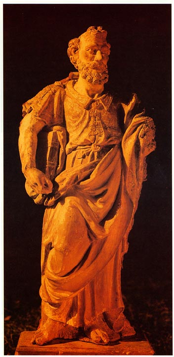 |
Apparent but misleading damage. This Apostle, from the end of the 16th, beginning of the 17th centuries, has a completely worm-eaten surface that appears to indicate that the sculpture is infected. The scanned cross-sections show that the damage is only superficial and localised in the left-hand side of the object. |
 |
|
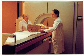
| The scanner used is that which is normally used in medicine. Here, an Apostle is placed by Dr Bensimon and Gilles Perrault on the table ready for scanning. The angle of the table and the scanner can be controlled by the computer console or the control unit under the table. | |
 |
|
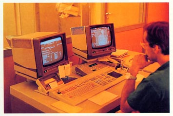
| A photo of the scanner control console. On the screen to the left is the programme chosen and, on the right, the scan of the Apostle with the scanned cross-sections. | |
 |
 |
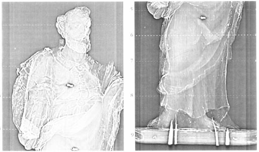
| An examination under a microscope of the various paint layers shows that they are different in the lower and upper parts of the Virgin; a subsequent observation by microscope after opening a window in the paint reveals the presence of suspect glued joints. As it was impossible to remove all of the paint, the scanner appeared to be the most appropriate solution. Various issues arose: what was the importance of the pieces that were added? Was there a cavity containing religious relics, hidden by a subsequent restoration, as frequently occurs for bases of this epoch? | Cross-section no. 4 proves that the Child Jesus was cut from the same block as the Virgin: the concentric rings concord perfectly, except for the two pieces subsequently added: one, upper right, corresponding to the hand and book of the child, the other on the right-hand side. |
| Restoration of a Virgin with Child.
The sketch opposite shows the CT scan cross-sections taken of Cantal’s Virgin with Child, which was apparently not the subject of any issue of authenticity. The scanner revealed that one-half of the sculpture was at least from the 19th century. For understandable reasons, we are not able to reproduce the photo of this statue. For information purposes, featured here is a virgin of the same type from the Louvre Museum, the authenticity of which is not in doubt. |
 |
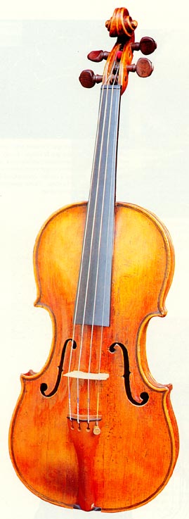 |
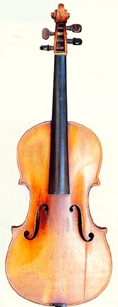
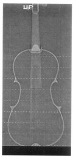
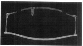
False notes in violins. These two violins were put through scanners for different reasons. The first, which is a fake Stradivarius produced by Jérôme Thibouville Lamy at the end of the 19th century, was the subject of an expert report by the expert, Mr Lanne. The scanner provided irrefutable proof of his conclusions, despite a misleading stamp. The second is, on the contrary, a violin from the 18th century; the scanned cross-section showed that it has been very skilfully restored. The photos from these two scanners constitute irrefutable proof in the event of a dispute in the courts. |
 |
 |
 |
| The overall scan of the fake Stradivarius identified the fraud, but not its curves or destiny. | The cross-section of the violin from the 18th century reveals its authenticity: the finesse of the assembly, the thicknesses of the wood, the work on the supporting bar, the arches used. The scanner shows the skilful work by the restorer: a second layer was added to the bottom via the interior and a crack was filled in the soundboard |
 |
 |
| Roman Virgin With cross-section no. 7, we discover various modern restorations to the chair, both to the edge of the sculpture and the interior, being the junction between the upper part of the statue, which is of that epoch, and all of the lower part, which was added recently. |
Cross-section no. 5 (photo above) supports this impression: the assembly and woodwork are not the same as those of authentic Stradivarius violins. |
![]() © Revue Experts
© Revue Experts